3h
Horse TNFa(Tumor Necrosis Factor Alpha) ELISA Kit
Horse TNFa(Tumor Necrosis Factor Alpha) ELISA Kit
tumor
500pg/mL
2.7pg/mL
Sandwich
7.8-500pg/mL
Cytokine;Tumor immunity;Infection immunity;
ELISA Enzyme-linked immunosorbent assays Code 90320007 SNOMED
DIF; TNF-A; TNFSF2; Cachectin; Tumor Necrosis Factor Ligand Superfamily Member 2
E05 478 566 350 170 or Enzyme-Linked Immunosorbent Assays,E05 478 566 350 170 or Enzyme-Linked Immunosorbent Assays,Horse (Equus ferus caballus) sera and plasma contain equine IgGs, Immunoglobulins. ELISA test are used to determine quantitatively the presence in horse serum of the antigen by a polyclonal antibody to the equine epitope selected for the ELISA kit. A blocking solution for the native horse or equine immunoglobulins in available in the ELISA protocol.
The TNFa(Tumor Necrosis Factor Alpha) ELISA Kit is a α- or alpha protein sometimes glycoprotein present in blood.Aplha, transcription related growth factors and stimulating factors or repressing nuclear factors are complex subunits of proteins involved in cell differentiation. Complex subunit associated factors are involved in hybridoma growth, Eosinohils, eritroid proliferation and derived from promotor binding stimulating subunits on the DNA binding complex. NFKB 105 subunit for example is a polypetide gene enhancer of genes in B cells.
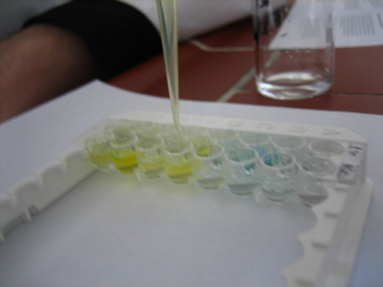
The test principle applied in this kit is Sandwich enzyme immunoassay. The microtiter plate provided in this kit has been pre-coated with an antibody specific to Tumor Necrosis Factor Alpha (TNFα). Standards or samples are then added to the appropriate microtiter plate wells with a biotin-conjugated antibody specific to Tumor Necrosis Factor Alpha (TNFα). Next, Avidin conjugated to Horseradish Peroxidase (HRP) is added to each microplate well and incubated. After TMB substrate solution is added, only those wells that contain Tumor Necrosis Factor Alpha (TNFα), biotin-conjugated antibody and enzyme-conjugated Avidin will exhibit a change in color. The enzyme-substrate reaction is terminated by the addition of sulphuric acid solution and the color change is measured spectrophotometrically at a wavelength of 450nm ± 10nm. The concentration of Tumor Necrosis Factor Alpha (TNFα) in the samples is then determined by comparing the O.D. of the samples to the standard curve.
